 Add as a preferred source on Google
Written by The Skin Firm clinical content team · Reviewed by Dr. Karishma Singh, BHMS, FCHD (Founder, Skin & Aesthetic Specialist) · Reviewed on 11 November 2026
Quick answer
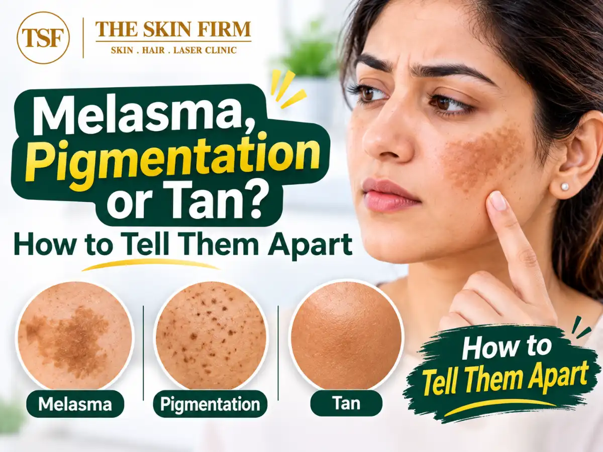
Melasma is one type of hyperpigmentation. It shows up as symmetrical, blotchy brown or grey-brown patches on both cheeks, the forehead, upper lip or jawline, and it tends to keep coming back. A tan is an even darkening of sun-exposed skin with sharp edges, and it fades within weeks once you avoid the sun. Other pigmentation, such as acne marks or sun spots, sits exactly where its trigger was. A doctor can confirm which one you have.
In Pune, many people call every dark patch "pigmentation" or "tan" and reach for a de-tan pack. That rarely helps when the patch is actually melasma, which behaves very differently on brown skin. This guide explains how the common types differ, how a doctor tells them apart, and a sensible first step for each. If you already suspect melasma, our page on pigmentation and melasma treatment in Pune explains how the clinic approaches it.
Is melasma the same as hyperpigmentation?
Hyperpigmentation is an umbrella term for skin that is darker than your usual tone. It happens when pigment cells make extra melanin, or when melanin settles deeper in the skin.
Melasma is one specific condition under that umbrella. Others include sun tan, post-inflammatory hyperpigmentation (PIH), freckles and sun spots. Each has a different cause, lasts a different length of time and needs different care. For a wider look, see our guide to the causes of skin pigmentation.
Melasma vs tan vs other pigmentation: comparison table
- Melasma: What it looks like: Symmetrical, blotchy light-brown to grey-brown patches, irregular edges; Usual location: Cheeks, forehead, nose, upper lip, jawline; Cause and triggers: Sun, visible light, heat, hormones, genes, irritating products; How long it lasts: Months to years; often returns each summer; What helps first: Tinted sunscreen, a hat, gentle skincare, a doctor's plan
- Sun tan: What it looks like: Even darkening with sharp lines at collar, sleeve or watch strap; Usual location: Any uncovered skin; Cause and triggers: UV exposure; How long it lasts: A few weeks to a couple of months; What helps first: Sunscreen, shade, covering up, time
- PIH: What it looks like: Flat brown to grey marks in the shape of an earlier spot or injury; Usual location: Sites of acne, eczema, bites, burns, waxing or threading; Cause and triggers: Inflammation or injury; How long it lasts: Several months; deeper marks a year or more; What helps first: Treat the cause, stop picking, sunscreen
- Freckles: What it looks like: Small light-brown dots, darker in summer, lighter in winter; Usual location: Nose, cheeks, shoulders; Cause and triggers: Genes plus sun; How long it lasts: Come and go with the seasons; What helps first: Sunscreen
- Lentigines (sun or age spots): What it looks like: Flat, well-defined brown spots, larger than freckles, present all year; Usual location: Face, backs of hands, forearms; Cause and triggers: Years of sun; more common after 40; How long it lasts: Stay unless treated; What helps first: Sunscreen; a doctor's check if one changes
- Dermatosis papulosa nigra (DPN): What it looks like: Tiny dark, smooth, raised bumps; Usual location: Cheekbones, around the eyes, neck; Cause and triggers: Inherited tendency; common in brown skin; How long it lasts: Stay, may increase with age; What helps first: Harmless; a doctor can remove them
- Acanthosis nigricans: What it looks like: Velvety, thickened dark skin; Usual location: Back of neck, underarms, groin; Cause and triggers: Insulin resistance, excess weight, PCOS, some medicines; How long it lasts: Until the cause is addressed; What helps first: Blood sugar check, lifestyle care
What does melasma look like?
Melasma usually appears as flat, light-brown to grey-brown patches with irregular, map-like borders. It almost always affects both sides of the face in mirror-image shapes. It does not itch, and skin texture stays normal.
DermNet describes three main facial patterns:
- Centrofacial: forehead, cheeks, nose, upper lip and chin. This is the most common.
- Malar: the cheeks and nose.
- Mandibular: along the jawline and chin.
An Indian multicentre study of 331 people found the centrofacial pattern in 42%, malar in 39% and a combined pattern in 14%. Mandibular melasma alone was rare. Women outnumbered men by about four to one.
Epidermal, dermal or mixed: why depth matters
Melasma pigment can sit in the outer layer of skin (epidermis), the deeper layer (dermis), or both.
- Epidermal: darker brown with well-defined edges. It usually responds better to treatment.
- Dermal: lighter brown to bluish-grey with hazy edges. It is usually slower to improve.
- Mixed: a blend of both. DermNet notes this is the most common type, and it typically improves partly.
Deeper pigment needs more patience and gentler handling. Aggressive procedures on deep melasma can inflame the skin and add new pigment.
What triggers melasma in Indian skin?
Melasma is most common in medium-brown skin, like that of many people in Pune. Common triggers include:
- Sun and visible light. UV levels stay high for much of the year in India. The AAD notes visible light can also worsen melasma, especially in darker skin. In the Indian study, only 35% of patients used sunscreen.
- Heat. Many dermatologists see patches darken with heat from cooking stoves or hot commutes, even without direct sun.
- Hormones. Pregnancy is a well-known trigger. In the Indian study, 51% of women with more than one pregnancy had melasma. Birth control pills can contribute, and thyroid problems are sometimes linked.
- Family history. About a third of Indian patients in that study had an affected relative.
- Irritating products. Fragranced creams, harsh scrubs, lemon packs and stinging "de-tan" products can inflame skin and deepen patches.
If you are pregnant or planning to be, our guide to pregnancy-safe skincare covers what to avoid.
Is this melasma or tan? A quick self-check
These clues help, but they do not replace a diagnosis.
- 1. Is it on both cheeks in a similar shape? Symmetry leans towards melasma.
- 2. Are there sharp lines at your collar, sleeve or watch strap? That is typical of a tan.
- 3. Did it fade after a few weeks indoors? Tan fades; melasma lingers or returns.
- 4. Did it start in pregnancy, on the pill or after a new product? Melasma is more likely.
- 5. Is the mark exactly where a pimple or rash was? That suggests PIH.
- 6. Is it velvety and thick, on your neck or underarms? Think of acanthosis nigricans.
You can have more than one at once, such as melasma with a tan over it. If yours looks like a simple tan, our guide to tan removal at home vs clinic treatment compares the options.
How do doctors diagnose melasma?
Melasma is usually diagnosed from your history and an examination. A doctor will typically:
- 1. Take a history: when it began, pregnancies, pills, thyroid issues, sun habits, products and family history.
- 2. Examine the pattern: symmetry, borders, colour and location.
- 3. Use a Wood's lamp: a handheld ultraviolet light used in a dim room. Shallow pigment looks more prominent under it; deep pigment does not.
- 4. Use dermoscopy: a magnified, polarised view of the skin. A brown, well-defined network suggests shallow pigment, while grey-blue, hazy patterns suggest deeper pigment.
- 5. Occasionally take a biopsy if the picture is unclear.
In a Goa study in the Indian Dermatology Online Journal, the two tools agreed only moderately on depth. The authors noted the Wood's lamp is less reliable on darker skin, so dermoscopy is often more useful for Indian skin. A doctor will also rule out look-alikes, such as drug-related pigmentation or naevus of Ota.
Why getting the diagnosis right matters
- Bleaching and "fairness" creams. Unsupervised steroid-containing creams can thin the skin and cause visible red vessels. In the Indian study, some previously treated patients had visible blood vessels (7%) or exogenous ochronosis (3%), a stubborn blue-grey darkening.
- Aggressive peels, scrubs and lasers. DermNet notes peels and lasers can worsen melasma or cause new PIH if not used cautiously.
- Missing a health signal. Treating acanthosis nigricans as "tan" can mean missing early insulin resistance or PCOS.
Please do not self-medicate with prescription creams, even if they helped a friend.
First steps for each type
Sun protection is the shared foundation for every type of pigmentation.
- For everyone: a broad-spectrum sunscreen of SPF 30 or higher, applied 15 minutes before going out and reapplied every two hours. Add a wide-brimmed hat, sunglasses and shade.
- Melasma: choose a tinted sunscreen with iron oxides, which also helps against visible light. Keep skincare fragrance-free, and see a doctor before starting active creams.
- Tan: protect your skin and give it time.
- PIH: get the acne or eczema under control first, and avoid picking.
- Freckles and lentigines: show a doctor any spot that grows, bleeds or changes colour.
- DPN: leave the bumps alone or have a doctor remove them. Never cut or burn them at home.
- Acanthosis nigricans: ask your doctor about a blood sugar test. Our article on dark neck and acanthosis nigricans explains more.
There is no cure for melasma, but it can often be controlled. Our upcoming guide, "Does melasma ever go away?", covers treatment and recurrence in detail (coming soon).
When to see a doctor
Book a skin check if:
- patches are spreading, or have not faded after two to three months of good sun protection;
- pigmentation started during pregnancy, on the pill or with a new medicine;
- a cream has made your skin red, thin or darker;
- a spot is raised, irregular, growing, bleeding or changing colour;
- you have velvety darkening with weight gain, irregular periods or a family history of diabetes.
You can book a consultation at The Skin Firm, a skin, hair and laser clinic at NIBM Road, Pune, to have your pigmentation assessed. A doctor can identify which type you have and discuss a realistic plan. Results vary from person to person.
References
- Melasma (facial pigmentation) — DermNet
- Melasma: Causes — American Academy of Dermatology
- Melasma: Self-care — American Academy of Dermatology
- A Cross-Sectional, Multicentric Clinico-Epidemiological Study of Melasma in India — Dermatology and Therapy (via PubMed Central)
- Comparison of Dermoscope and Woods Lamp as A Tool to Study Melanin Depth in Melasma — Indian Dermatology Online Journal (via PubMed Central)
This article is for general information and isn't a substitute for a medical consultation. Editorial note: drafted with AI assistance and medically reviewed before publication.